
Shoulder Arthroscopy
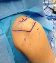
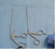
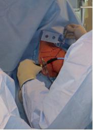
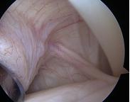
Arthroscopy is a minimally invasive way to perform shoulder surgery. To perform an arthroscopy Dr. Chalmers will make a small incision 5 millimeters (1/5th inch) and insert a small fiberoptic camera into the shoulder joint. Once the joint is filled with saline fluid, the entire shoulder joint can be visualized using the camera. Secondary incisions can be made to insert long thin instruments made specifically for arthroscopy. If you have a shoulder arthroscopy, initially after surgery the shoulder will be very swollen with fluid used to visualize the joint during the surgery. This fluid dissipates rapidly, but the shoulder may remain swollen because of the procedure performed.
Many procedures that used to require an open incision can now be performed through an arthroscopy. Examples include removal of inflammation, removal of bone spurs, treatment of tendonitis, rotator cuff repair, labral repair, and shoulder stabilization after a dislocation. Because the procedure involves small incisions and minimal damage to the deltoid muscle, many surgeons believe patients that undergo arthroscopy may have less post-operative pain, less postoperative stiffness, fewer post-operative infections, and an overall decreased rate of complications. Most shoulder arthroscopies are performed on an outpatient basis, which means patients can have their procedure at a surgery center instead of the hospital and can go home the day of surgery instead of staying the night in the hospital.
Not all procedures in the shoulder can or should be performed through an arthroscopy. For instance, shoulder fractures, shoulder replacements, and revision of prior surgery often requires a traditional incision in the front of the shoulder.
Click here to download the PDF.
You will need the Adobe Reader to view and print these documents